COVID-19 disease, caused by the coronavirus and which has as principal form of remedy social isolation, has played a great role in the intense use of the most diverse formats of technology.
Doctors in the area have come together to fight this great evil, and an example of this is the partnership between Medical Harbor, Dr. Jezreel and Dr. Omir Antunes Paiva from RAIOSS.
As already mentioned in the post Teleradiology: 7 benefits for medical professionals and patients Teleradiology, or telemedicine offers a series of advantages for professionals in the medical field. More than ever, the gift of technology has been present in the medical routine so that it is possible to continue medical education, health treatments and research in the area.
Partnership to save lives
With the creation of the coronacases.org platform, developed by Dr. Omir and RAIOSS, it was possible to gather a series of radiographic images of patients affected by COVID-19.
Using our platform for educational medical imaging, Athena Hub, Dr. Jezreel of the Radiology with Jezreel YouTube channel, made a didactic and accurate analysis of the consequences of coronavirus in the human body systems.
Case analysis with Dr. Jezreel
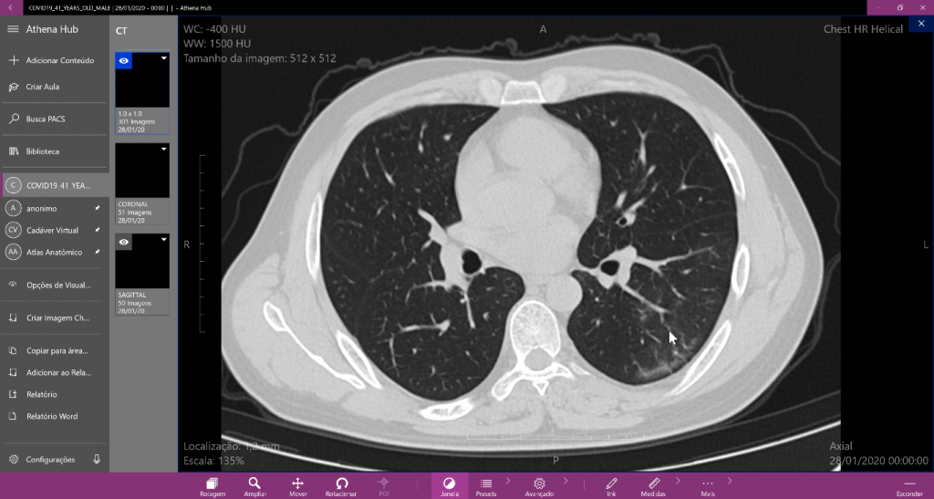
The patient in question is 41 years old and Dr. Jezreel points out that:
“It is possible to find the faint ground-glass opacities in the posterior region of the lower side of the left lung. It(the disease) pulps the subpleural region a little and has some parenchymal bands. ”
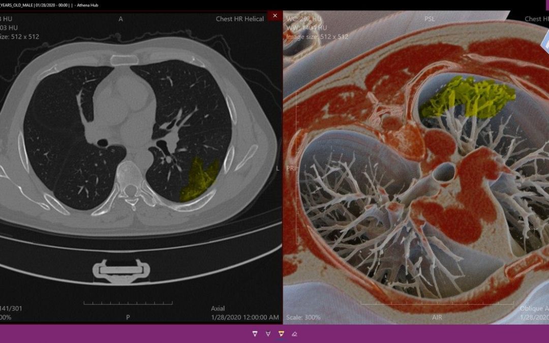
Posterior region of the lower side of the left lung.
To make this observation, Dr. Jezreel uses the scroll and zoom tools, which allow for a clear and fluid view of the image.
In the Sagittal MPR view, it is possible to better visualize the similarity bands and the opacity of the frosted glass in the lung region. According to Dr.’s observations, it is possible to state that part of the lung region can resist the disease.
“In the apical segment on the lower side of the left lung, you can see sparing in the subpleural region”.
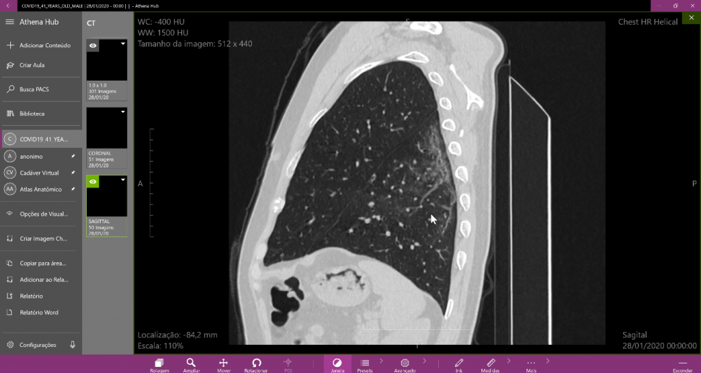
Sagittal visualization of the main imaging. One of many possible views in Athena Hub.
Finally, Dr. Jezreel uses the 3D Photorealism mode, which allows the reconstruction of medical imaging in high fidelity. With it, it is possible to make a more in-depth realistic analysis of the disease.
With the mode activated it is possible to quickly identify the situation of the patient’s respiratory system.
“The reconstruction with the photorealism mode clearly shows the distribution of opacity. See how there is a subpleural area spared in these cases. ”

Photorealism mode, that uses Photorealistic techniques to reconstruct medical imaging.
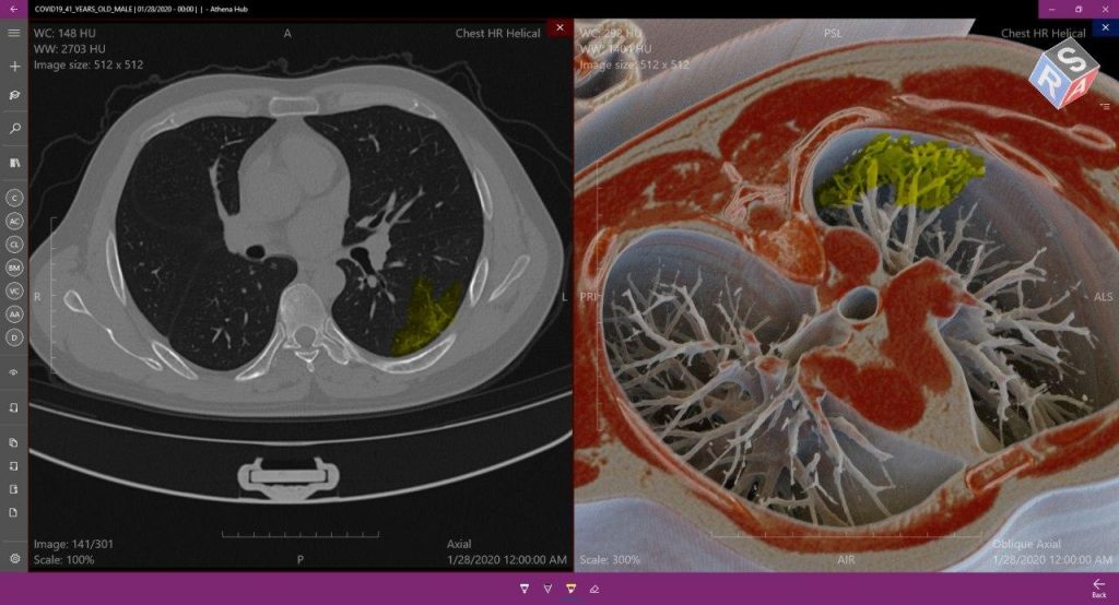
Comparative visualization of COVID-19, possible because of the multi-series feature.
It is possible to observe how the tools zoom, rotate and windowing work in harmony and precision. The high response rate in the movement of the imaging allows accurate and rapid diagnosis, assisting in the combat against COVID-19.
Given the rapid development of the situation, it is important to know how to identify the disease in medical imaging, all the while studying it to fight it.
With that in mind, Medical Harbor is making Athena Hub available for free for a limited time so that medical professors and students can continue to improve their knowledge during the quarantine period. To learn more about the subject, click here.
Be sure to follow us on social media to stay on top of relevant updates on the subject!