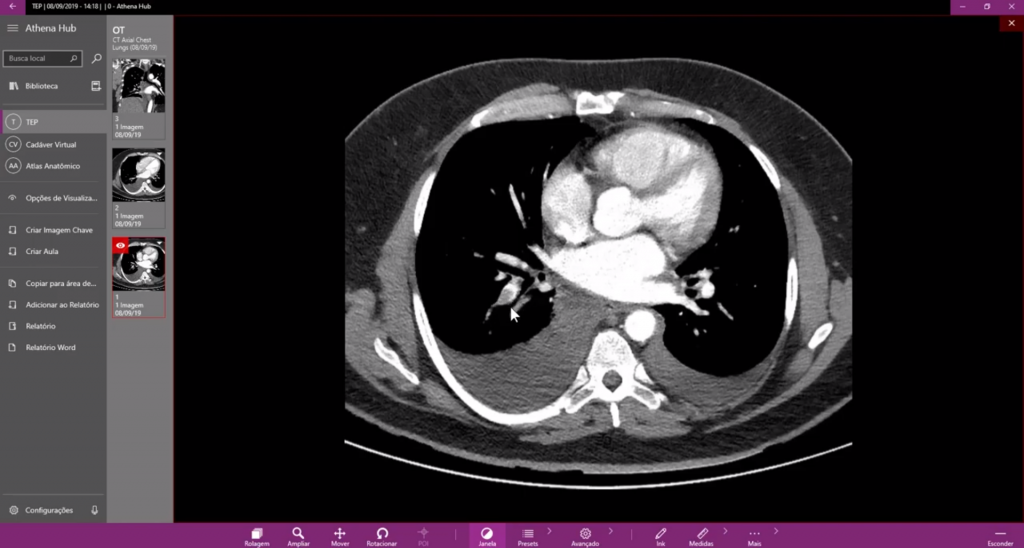
Pulmonary embolism is a disease consisting of the formation of clots inside veins located on the lower members, usually in the calves and making their way to the pulmonary arteries, causing a pulmonary embolism. The signs of the disease can occur in many ways and it can be discovered with the help of a DICOM Viewer.
Sometimes you must wonder: How can I be sure that a DICOM Viewer is useful on a day to day basis? A DICOM Viewer, for instance, can be used to diagnosis and make a treatment plan for Pulmonary embolism. The youtube Channel Radiology with Jezreel approached the subject and this time, the highlighted software, used by Dr. Jezreel is the Athena Hub, one of our solutions focused on medical education, and acting as a great form of checking out the multiple functions of a DICOM Viewer.
All tools and observations made during the video can be reproduced in our DICOM Viewers, Athena Athena DICOM Expert, and Athena DICOM Essential. This is a great opportunity to know a little bit more about the software in action.
How can we identify the Pulmonary embolism?
As said by Dr. Jezreel the fastest and most practical way of identifying Pulmonary embolism is thought a Computed Angiotomography of the pulmonary arteries, a commonly requested exam when Pulmonary embolism is suspected.
Image Analysis
While opening the exam into Athena, Dr. Jezreel explains that:
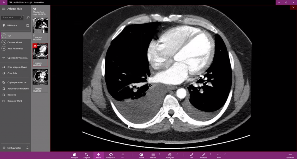
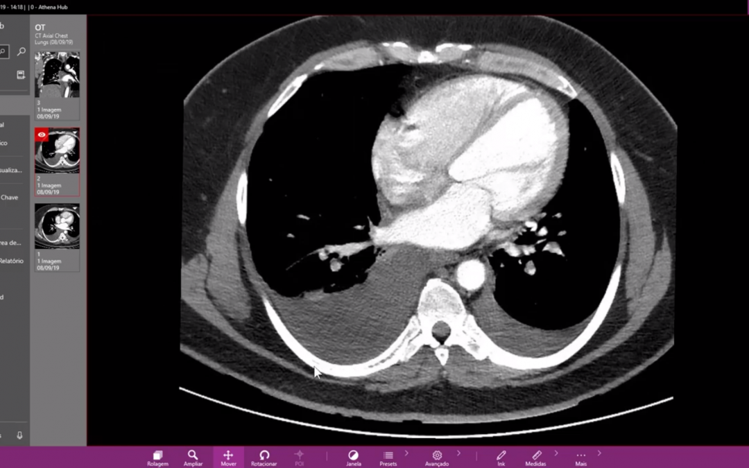
“ We have a failure while filling one of the pulmonary artery branches. This failure in the felling represents a thrombus. Observe that it won’t let the contrast fill the vase completely. Compare with this other arterial branch that is completely full of contrast.”
Besides, the zoom and moving tools facilitate the evaluation of the area of interest, allowing a more precise observation of the failed filled branches of the pulmonary artery. The tools can be reached easily by using shortcuts, guaranteeing a rate, and high productivity.

It is also possible to make a comparison between the pulmonary vases on the left and right sides. Dr. Jezreel explains that:
“[…] We have a bigger bilateral pleural effusion on the right, but we also have a little on the left too.”
Still analyzing the same study but now in another angle, in a coronal reconstruction, it is possible to observe the failed fillings in many pulmonary artery branches. The fastness and precision of the DICOM Viewer facilitate the detection of the failed fillings and take all the doubts of any other possible diagnosis.

It is also important to emphasize that in only 3 minutes, it was possible to diagnose and observe the behavior of a pulmonary embolism, in distinctive reconstruction plans and with only two manipulation tools, in the same DICOM Viewer.
The application of a DICOM Viewer goes on beyond the presented here and can be used for other means.
The Athena DICOM Expert, for instance, possesses advanced reconstruction tools, and others such as MIP, AIP, MinIP, that can assist in specific diagnoses.
The Athena DICOM Essential is directed towards professionals that are looking for a DICOM Viewer that can make fast diagnoses, using only basic tools.
Check out our comparative features charts and find out which is the best DICOM Viewer for your needs.
But, if your demands are others, such as, creating classes and using a DICOM Viewer for educational purposes, find out more about the Athena Hub, our solution made for education and that carries specific tools for it.
For any purposes whatsoever, be it diagnoses, studies, planning classes or surgeries, curiosity or just to find out about the functionality of a DICOM Viewer, the Athena family has the ideal version for you! If you have any doubts, contact us!
To see the complete video with Dr. Jezreel access the link:https://www.youtube.com/watch?v=koX0ynzypVY

